What exactly happens during a slit-lamp examination?
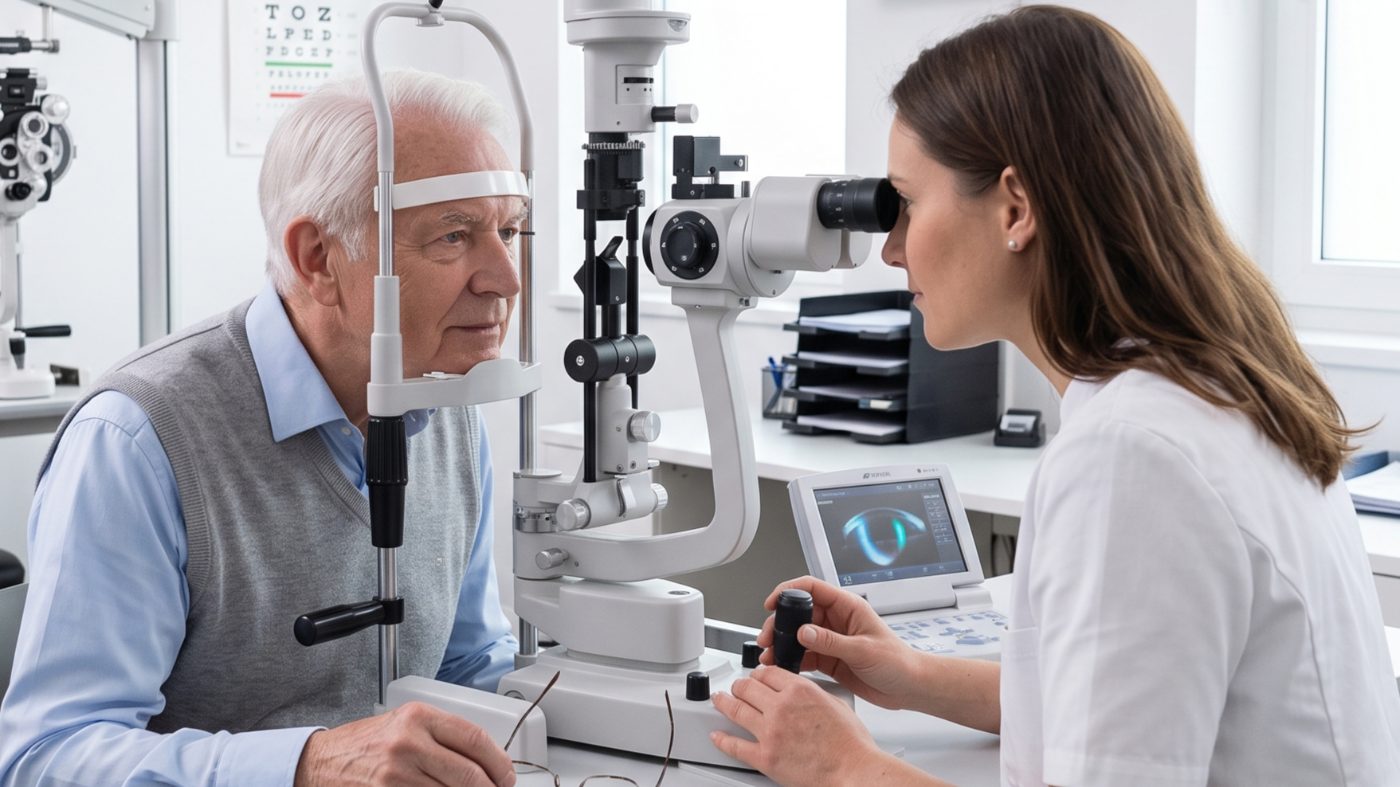
The slit-lamp examination is the most reliable method for detecting cataract (a clouding of the eye’s natural lens). A special microscope with a narrow, bright band of light makes the lens of the eye visible and shows whether and where it is clouded. The examination takes only a few minutes and is painless.
This article explains step by step what to expect at this appointment and why the examination is considered the gold standard.
What is a slit lamp in the first place?
A slit lamp is a binocular microscope combined with a swivelling light source. The beam of light can be narrowed to a thin slit, hence the name. This narrow slit of light cuts optically through the transparent structures of the eye, rather like a CT cross-section, only with light instead of X-rays.
This allows the cornea, anterior chamber, iris and lens to be assessed layer by layer. Cloudy areas in the lens show up as grey or yellowish patches whose position and extent can be determined precisely.
How the examination proceeds
1. Preparation You sit down in front of the device and rest your chin and forehead on a support. This keeps your head still, much like at the optician.
2. First inspection without drops The ophthalmologist first inspects the eyelids, conjunctiva and cornea with the pupil at its normal size. Some lens opacities can already be seen at this stage, especially central ones.
3. Pupil dilation (mydriasis) For a full assessment of the lens out to its edge, eye drops are usually given to widen the pupil. These drops need 20 to 30 minutes to take effect. During this time you can take a seat in the waiting area.
4. Examination with the pupil dilated With the pupil wide, the ophthalmologist sees the entire lens, including the outer zones where opacities often form first. He or she assesses the position, density and shape of the clouding and assigns it to a particular type of cataract.
5. Additional measurements Directly afterwards, the eye pressure (intraocular pressure) is often measured and the back of the eye is checked, to rule out other conditions such as glaucoma or retinal changes.
What the ophthalmologist actually sees
At the slit lamp, cataract shows itself in different patterns, depending on which part of the lens is affected:
- Nuclear cataract: yellowish to brownish clouding in the centre of the lens, often slowly progressive.
- Cortical cataract: spoke-shaped, whitish opacities at the edge of the lens.
- Posterior subcapsular cataract: disc-shaped clouding directly in front of the back surface of the lens, often causing severe glare early on.
This classification helps not only with the diagnosis but also with judging how much the clouding already impairs vision and how it is likely to develop.
An example from practice: with a nuclear cataract, patients often first notice a yellowish tinge to their vision. The difference frequently only becomes obvious when compared with the other, still clear eye.
Biometry: the next step after the diagnosis
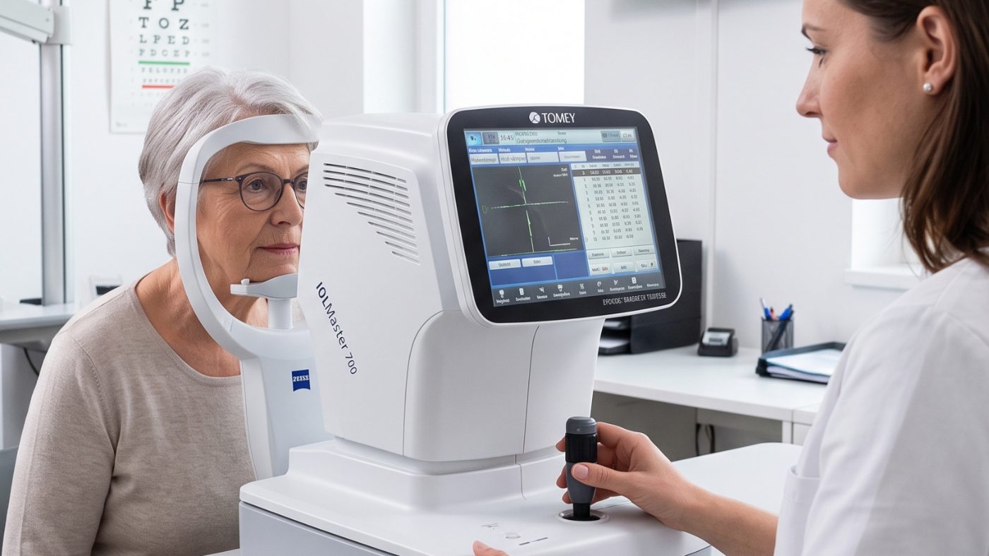
If the slit-lamp examination confirms a cataract and you wish to have surgery, a further appointment usually follows: biometry (measuring the eye). It is a separate measurement and differs from the slit-lamp image.
An optical measuring device, often referred to as an IOL Master, records the axial length of the eye, the curvature of the cornea and the depth of the anterior chamber. These values feed into a calculation formula that determines the right power for the artificial lens.
With a very dense clouding, the optical method sometimes no longer delivers a reliable result. In that case, ultrasound biometry is used, which measures the same values by a different route.
Biometry itself is likewise painless and takes only a few minutes per eye. Unlike the slit-lamp examination, it does not require pupil dilation. The two examinations complement each other: the slit lamp establishes the diagnosis, biometry prepares for the operation.
Depending on the practice, both appointments can take place on the same day. More often, however, a few days to a few weeks lie between them, leaving enough time for evaluating the results and choosing the lens.
Is the examination uncomfortable?
No. The slit lamp itself does not touch the eye. At most, the brightness of the light can be briefly unpleasant. After pupil dilation, the eyes are more sensitive to light for a few hours and near vision is blurrier. Sunglasses for the way home are therefore helpful. Driving is usually not recommended during this period.
Frequently asked questions
Do I need to fast before a slit-lamp examination? No, that is not required. You can eat and drink normally beforehand.
How long does the whole examination take, including pupil dilation? Allow 45 to 60 minutes, since the drops need time to take effect. The actual inspection at the slit lamp takes only a few minutes.
Can I drive myself home after the examination? Directly after pupil dilation, near vision and vision in bright light are impaired. Many practices advise arranging a lift or using public transport to get home.
Is a slit-lamp examination enough to plan an operation? For the diagnosis, yes. Before a planned operation, additional measurements such as biometry are carried out to calculate the right artificial lens.
Are other eye conditions also detected during a slit-lamp examination? Yes. Besides cataract, the slit lamp can also reveal corneal changes, signs of inflammation of the iris or abnormalities of the anterior chamber. Combined with the eye pressure measurement, the ophthalmologist also largely rules out glaucoma.
What is an endothelial cell count and is it always done? This additional measurement records the number of cells in the innermost layer of the cornea. It is not part of the routine, but is used specifically when a corneal condition such as Fuchs’ dystrophy is suspected.
Next steps
The slit-lamp examination is usually the first step on the way to a possible operation. You can find out what further measurements follow on our page about the preliminary examination. If you are unsure whether your symptoms point to cataract, also read Do I have cataract?.
Sources
- Grauer Star - DocCheck Flexikon
- Grauer Star - Österreichische Ophthalmologische Gesellschaft
- Grauer Star: Diagnose und Behandlung - Augenarzt Online
- Ablauf der Spaltlampenuntersuchung - American Academy of Ophthalmology
- Grauer Star (Katarakt): Diagnose mit der Spaltlampe - Öffentliches Gesundheitsportal Österreichs